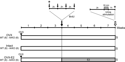
Figure 2.
Experimental design for assessing cell survival and the activation of newborn neurons in the MOB and AOB. Three additional groups of adult WT and ArKO female mice were submitted to 3 distinct hormonal treatments for 3 wk. OVX and OVX-E2 groups were ovariectomized on the first day and returned to their home cages for 3 wk. On d 21, all mice received 4 BrdU injections (at 2-h intervals) and were returned to their respective cages for 4 wk. OVX-E2 mice received implants the day after BrdU injections and kept the implants for the remaining 4 wk of the experiment. Vaginal smears (stars) were performed in intact mice at the time of the first BrdU injection on d 21 and again on the final day of the experiment, just before sacrifice (S). On the final day, all mice were separated into 2 subgroups, isolated for 2 h before stimulation, and exposed for 30 min to either deionized water or to male urinary odors. Mice were killed 60 min later to assess immediate Zif268 gene expression.

