Figure 3.
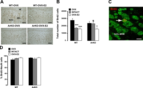
Influence of estradiol on the survival of newborn neurons in the adult MOB. A) Representative images showing newly generated BrdU cells in the GrCL of the MOB, analyzed 4 wk after BrdU injections. Number of BrdU cells was decreased in WT-OVX-E2 and ArKO-OVX-E2 compared with WT-OVX and ArKO-OVX female mice. SEL, subependymal layer; Mi, mitral cell layer. B) Total number of BrdU+ cells in the MOB GrCL from the 6 experimental groups. C) Representative image of BrdU-NeuN double-labeled cell (arrow) under confocal microscopy. D) Percentage of BrdU-NeuN double-labeled cells counted in the GrCL of the MOB. OVX: WT, n = 6; ArKO, n = 6. Intact: WT, n = 4; ArKO, n = 4. OVX-E2: WT, n = 6; ArKO, n = 5. Values are expressed as means ± se. Scale bars = 60 μm (A); 6 μm (C). *P < 0.05, ***P < 0.005 vs. OVX; SP < 0.05 vs. WT; 2-way ANOVA followed by Fisher's post hoc test.

