Figure 4.
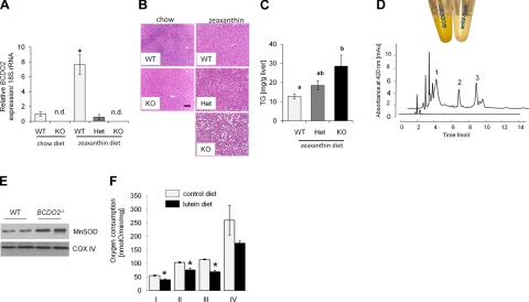
Carotenoid accumulation impairs mitochondrial function. A) Expression of BCDO2 mRNA in the liver of WT, BCDO2-HET, and BCDO2-KO mice subjected to different dietary interventions. Values represent means ± sd from 5 mice/group. n.d., not detectable. *P ≤ 0.05. B) Representative liver sections (×20) of WT, HET, and KO mice subjected to different dietary interventions. C) Liver triacylgylceride levels of WT, HET, and KO mice subjected to zeaxanthin diet. Values not sharing a common letter (a, b) are statistically different (P<0.05); 1-way ANOVA and LSD post hoc comparison. D) Colors of isolated hepatic mitochondria of BCDO2−/− and WT mice fed a diet supplemented with diet. HPLC trace at 420 nm of a lipophilic extract of isolated hepatic WT (bottom trace) and BDCO2-deficient (top trace) mitochondria. Peaks 1–3 show spectral characteristics identical to those of 3-dehyrolutein derivatives depicted in Fig. 2C. E) Immunoblot analysis for MnSOD with protein extracts from hepatic mitochondria (50 μg protein/lane). Staining for COXIV was used as a loading control. F) Oxygen consumption of complexes I–IV in isolated hepatic mitochondria from age-matched male BCDO2−/− mice fed lutein diet vs. control diet. Oxygen consumption was measured by the addition of complexes I–IV (see Materials and Methods) in the presence of 100 mM ADP to determine maximal ADP-dependent respiration rates. *P ≤ 0.05.

