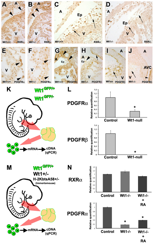
Fig. 4.
Status of RA-related receptors in Wt1-null epicardial cells. (A-D) RXRα immunoreactivity in E11.5 wild-type (A) and Wt1-null (B) embryonic epicardium is not significantly different (arrowheads); differences in RXRα epicardial immunoreactivity in E13.5 wild-type (C) and Wt1-null (D) embryos are evident (arrows). (E,F) PDGFRα immunoreactivity in the ventricular epicardium of E11.5 Wt1-null embryos (F, arrowheads) is reduced when compared with stage-matched wild types (E, arrowheads). (G-J) PDGFRβ immunoreactivity is reduced in the ventricular (H, arrowhead) but not atrial epicardium (H, arrow) of E11.5 and E13.5 Wt1-null embryos (J, arrow) when compared with E11.5 (G, arrowheads) and E13.5 (I, arrow) wild types. The Wt1-negative cells of endocardial cushions are PDGFRβ positive (J, arrowhead). (K) Procedure for the characterisation of embryonic epicardial cells. Wt1GFP/+ and Wt1GFP/- hearts were dissected, GFP-positive epicardial cells sorted, mRNA extracted and analysed by qPCR (L). (M) Procedure for the isolation and qPCR analysis of control (Wt1GFP/+) and Wt1-null (Wt1GFP/−) immortalised epicardial cells. (N) qPCR analysis of Rxra, Pdgfra. RA treatment (1 μM RA for 24 hours) of mutant cells did not affect Rxra levels but did increase Pdgfra levels in Wt1-null cells. All asterisks indicate statistical significance (P<0.05, 10 d.o.f.). Data are mean+s.e.m. Abbreviations: A, atrium; Ep, epicardium; V, ventricle.

