Fig. 6.
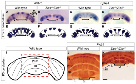
Embryonic and neonatal mispatterning of Zic mutant cerebella correlates with adult foliation defects. (A-D) Anterior views of in situ hybridization for Wnt7b (A,B) and Epha4 (C,D) in whole E17.5 cerebella. Wild-type parasagittal stripe patterns (A,C). Anterior stripes were reduced in size or absent in Zic1−/−;Zic4−/− cerebella (B,D). d, dorsal; v, ventral; a, anterior; p, posterior; m, medial; l, lateral. (E-H) Diagrams depict normal (E,G) and disrupted expression patterns (F,H). Regions of stripe differences are indicated by asterisks and brackets in A-H. (I) The anterior surface of a P3 cerebellum with vermis highlighted (red dotted box). (J,K) Anterior vermis views of anti-Plcβ4 whole-mount antibody staining of P3 cerebella. Wild-type cerebellar vermis (J) with four major parasagittal stripes of expression (brackets; M, medial; L, lateral) perpendicular to folia (roman numerals). Fissures (dotted lines in J and K) excluded antibody penetration and produced areas with no visible staining between lobes. Zic1−/−;Zic4−/− (K) cerebella lacked one anterior lobule and remaining stripes were abnormally patterned. The medial stripes of lobe II/III were relatively unaffected, but the lateral stripes were much narrower compared with wild type. Lobe IV/V patterns were completely disrupted with irregularly shaped and bilaterally asymmetric regions of expression. Scale bars: 500 μm in A-D; 250 μm in J,K.

