
Figure 6.
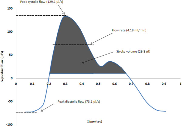
Example of MRI flow waveforms in the cerebral aqueduct. A typical MRI-derived flow waveform, demonstrating the possible measures extracted for quantification. Stroke volume is the most common parameter used, and is a measure of the net flow through the vessel/region of interest, in one direction (i.e., over approximately half the cardiac cycle). Flow rate has also been used frequently, and is the mean flow rate for flow in one direction. Peak flow is used less frequently, and is a measure of the highest (i.e., systolic, or lowest for diastolic) flow rate over the entire cardiac cycle.