
Figure 6.
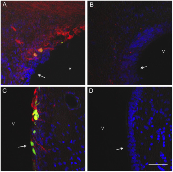
Nestin immunoreactivity is decreased in the subependymal zone following Ecrg4 over-expression. A and C: Nestin immunoreactivity in rat subependymal zone. When examined under low (Panel A, 200×) or higher (Panel C, 400×) magnification, nestin immunoreactivity labeled neuroepithelial progenitor cells that were present and proliferating after the i.c.v. injection of AdGFP into injured rat brains. Nestin was also detectable in the ependymal cell layer as denoted by arrows in the ventricle (v) where it co-localized with GFP positive cells. Blue = DAPI, Red = nestin, green and yellow = GFP in AdGFP positive cells. B and D: Nestin immunoreactivity decreased in subependyma after ADEcrg4 injection. There was decreased nestin staining that was readily observed in the subventricular zone of AdEcrg4 treated rat brains when examined under low (Panel B, 200×) or higher (Panel D, 400×) magnification. These are the same zones where BrdU incorporation was also decreased (see Figure 5). The ependymal cell layer is denoted by arrows in ventricle (v). Blue = DAPI, Red = nestin Scale bar = 20 μm.