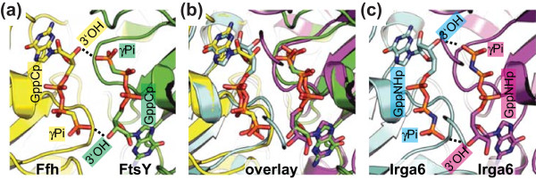
Figure 3.
Construction of the Irga6 dimer model. Views of the nucleotide-binding regions involved in formation of the dimers. (a) Crystal structure of the Ffh (yellow) FtsY (green) heterodimer (PDB 1RJ9) [20]. (b) Two molecules (cyan and magenta) of Irga6-M173A (PDB 1TQ6) [14] were adjusted to the Ffh-FtsY heterodimer, to give the best overlay for the bound nucleotides. (c) The model of the Irga6 dimer is shown. The trans interactions of the 3'OHs with the γ-phosphates are represented as dotted lines.

