
Figure 6. Fasciculation of sensory and motor projections is affected after ablation of Npn-1 from sensory neurons.
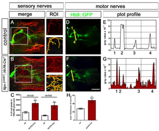
Defasciculation of cutaneous sensory nerves was assessed by calculating the number of neurofilament positive red pixels in a given 100×100 pixel region of interest (ROI). When compared to control embryos (A) this number is significantly increased in Npn-1cond −/− ;Ht-PA-Cre+ mutant embryos (B, C) for the cutaneous innervation of the dorsal and ventral limb (p dorsal<0.005, p ventral<0.005). Defasciculation of motor nerves was assessed by calculating a plot profile of Hb9::eGFP positive motor projections crossing a virtual line (1 = branch of n. radialis, 2 = n. radialis, 3 = n. medianus, 4 = n. ulnaris, arrowhead = n. musculocutaneous). When compared to control embryos (D, E) all four nerve branches can be found in Npn-1cond −/− ;Ht-PA-Cre+ mutant embryos, however, at slightly inappropriate positions to each other, and defasciculated fibers can be found in between major nerve branches. Quantification of the defasciculation of motor projections by measuring the thickness of the four major nerves revealed a significant increase in Npn-1cond −/− ;Ht-PA-Cre+ mutant embryos (H, 47.9±5.98 SEM) when compared to wt embryos (27.8±1.4 SEM, p<0.0001. The scale bar in (F) equals 100 µm for (A, B) and 400 µm for (D, F). n = 3 for all both genotypes; both limbs were quantified.