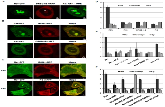
Figure 4. The RCN, DRBD1/2 and RG domains alter the subcellular localization of the HIV-1 Rev protein.
A) Subcellular localization of Rev in cells transfected with pRev-GFP alone (left panel) or with pRev-GFP in the presence of the RRE (right panel), and subcellular localization of pDRBD1/2-mRFP alone (middle panel). The cells were fixed 24 h post-transfection and the fluorescence (green or red respectively from Rev or DRBD1/2) was registered by confocal microscopy. Rev-GFP alone is visible exclusively in nucleoli (Nu); in the presence of RRE, Rev-GFP is also visible in the cytoplasm; pDRBD1/2-mRFP is visible in the cytoplasm and nucleolus. For RCN and RG a similar subcellular localization as for DRBD1/2 was observed (results not shown). B) Subcellular localization of Rev in the presence of RCN and DRBD1/2. HeLa cells were co-transfected with pRev-GFP, with pRCN-mRFP or with pDRBD1/2 and 24 h later, the respective fluorescence was observed. Green and red signals are illustrated alone and merged in the same cell. The yellow color in the merge indicates co-localization in nucleoli and cytoplasm. A similar result was observed for the RG domain (results not shown). C) In the presence of the RRE, RCN and RG drastically alter the subcellular localization of Rev. HeLa cells were transfected as described in B, but in addition with pCMVGag2RRE (to obtain RRE), and 24 h later the cells were observed by confocal microscopy. As illustrated for RCN-mRFP (upper panel), Rev-GFP is almost excluded from the nucleoli. At low magnification, a group of cells (lower panel) illustrates the variability of the distribution of Rev in the presence of RG and RRE. A similar result was obtained for DRBD1/2 (results not shown). D) Signal intensity was quantified in different cells as illustrated in A. GFP or red fluorescence either in the nucleolus (Nu), in the nucleoplasm (Nucleopl) or in the cytoplasm (Cy) were determined for each cell. E) Signal intensity was quantified in different cells as in B, as described in D. F) Signal intensity was quantified in different cells as in C, as described in D. As control HeLa cells were co-transfected with pRev-GFP and pmRFP, but no effect on Rev localization was detected (results not shown).

