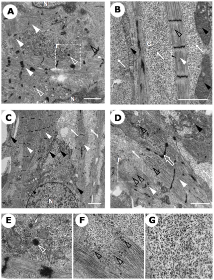
Figure 5. Ultrastructural analysis of human iPSC-derived cardiomyocytes.
Transmission electron microscopic images of beating colonies. Myofibrils with Z-bands (white closed arrowheads in A–D.), mitochondria (black closed arrowheads in B–D.), intercalated disk-like structure with desmosome (white open arrow in D.), atrial sercetory granules (electron-dense granules surrounded by double membranes. White open arrowheads in A. and E. (magnified image of A.)), glycogen granules (electron-dense small granules. Black open arrowheads in D. and F. (magnified image of D.)), ribosomal granules (electron-lucent small granules. White arrows in B–D. and G. (magnified image of B.)). N: nucleus. Scale bar = 2 µm, direct magnify, ×3000 (A), ×7000 (B), ×4000 (C), ×5000 (D).

