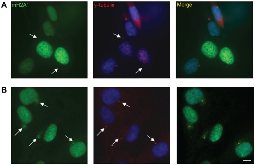
Figure 1. GFP fused macroH2A is not localized to the centrosome.
A. WI-38 cells were transfected with GFP-macroH2A1 and immune-stained for γ-Tubulin as a marker of the centrosome (Red). B. WI-38 cells were co-stained for γ-Tubulin (red) and macroH2A1-NHR antibody (Green). DNA stained with DAPI (Blue). Bar indicates scale of 10 µm.

