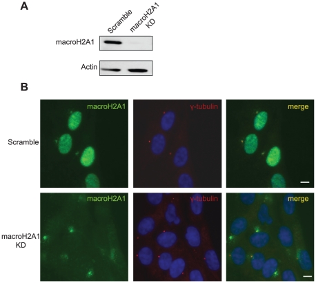
Figure 3. MacroH2A1-NHR antibody shows centrosomal staining in macroH2A1 KD cells.
A. Western blot verifying KD efficiency. B. WI-38 were transduced with either a scrambled vector or a macroH2A1 KD Cells were then subjected to immunofluorescence using antibodies against γ-Tubulin (red) and macroH2A1-NHR (green). DNA stained with DAPI (Blue). Bar indicates scale of 10 µm.

