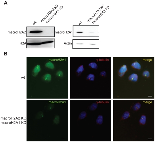
Figure 4. MacroH2A1-NHR antibody shows centrosomal staining in macroH2A1 and 2 double deficient cells.
To generate double deficient cells macroH2A1 was knocked down in macroH2A2 KO mESCs using stable shRNA lentiviral transduction A. Western blot analysis confirming absence of macroH2A2, using affinity purified macroH2A2 antibody (left panel), and macroH2A1, using macroH2A1-NHR (right Panel) in the targeted cells compared to control. B. Wt ESCs and macroH2A1 and 2 double deficient cells were stained with antibodies against γ-tubulin (red) and macroH2A1-NHR (green). Nuclei were stained with DAPI (Blue). Bar indicates scale of 10 µm.

