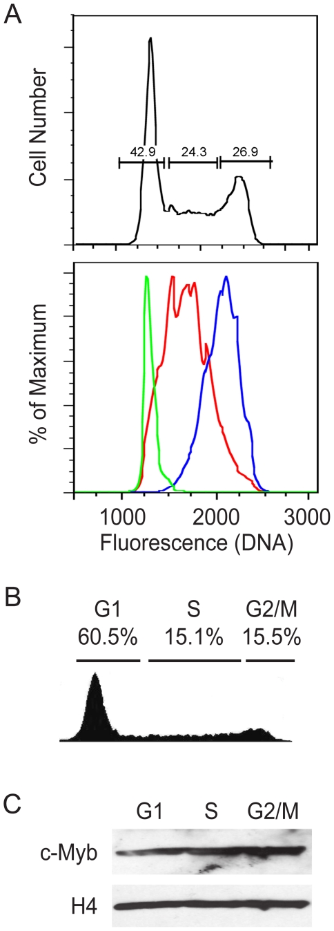
Figure 2. Chromatin-associated levels of c-Myb remain constant during the cell cycle.
(A) Cell cycle sorting of formaldehyde fixed cells. Asynchronously growing Jurkat cells were fixed with formaldehyde, stained with Hoechst 33342 then subjected to cell sorting, using a sorter equipped with a UV laser. The top panel shows the original cell cycle distribution and the gates that were used to collect the three cell cycle fractions. The lower panel shows the results of re-analyzing the cells collected in each sorted fraction. (B) Jurkat cell cycle analysis. Cell cycle histogram of a representative culture of Jurkat T cells progressing through the cell cycle. The cells were fixed with formaldehyde, stained with Hoechst 33342 and analyzed by flow cytometry. The fraction of cells in each cell cycle phase is indicated at the top. (C) Expression of c-Myb. Jurkat cells were fixed, stained with Hoechst 33342 and sorted into G1, S or G2/M fractions. Total chromatin was prepared from equal numbers of cells in each cell cycle fraction, then the cross-links were reversed and the samples were analyzed by Western blot (D) for expression of c-Myb protein. The blot was stripped and re-probed for histone H4 as a loading control. Note: The experiments in this figure were performed at least twice. The results shown are from a single experiment but are representative of all the trials.

