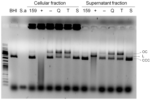
Figure 1. In vitro nuclease assays.
Plasmid DNA (100 ng, pTZ18R) was incubated at 37°C for 1 h with the cellular or supernatant fractions of various S. mutans strains, followed by electrophoresis on an agarose gel. Positive controls: S. aureus (S.a.) and UA159/pVE8009 (+). Negative controls: fresh BHI medium (BHI), cultures from UA159 (159) and UA159/pVE8010 (−). Q, T, S: UA159 derivatives containing LevQ-ΔSPNuc, LevT-ΔSPNuc and LevS-ΔSPNuc fusions, respectively. Open circular (OC), linear (L) and super-coiled (CCC) forms of the plasmid are labeled.

