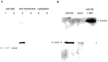
Figure 2. Western blots of LevQ protein generated using rabbit anti-LevQ antiserum.
(A) Various fractions of T/ldh and ΔlevQ culture were prepared from cells growing exponentially in BHI medium. 1, 3, 5: T/ldh; 2, 4, 6: ΔlevQ. An asterisk indicates the non-LevQ immune-reactive band in cell wall preparations. (B) Whole-cell lysates were prepared by bead-beating with 5% SDS using cells of UA159, a ΔlevQ mutant or UA159 treated with BS3. Both monomer and conjugates of LevQ (LevQc) are indicated by arrows.

