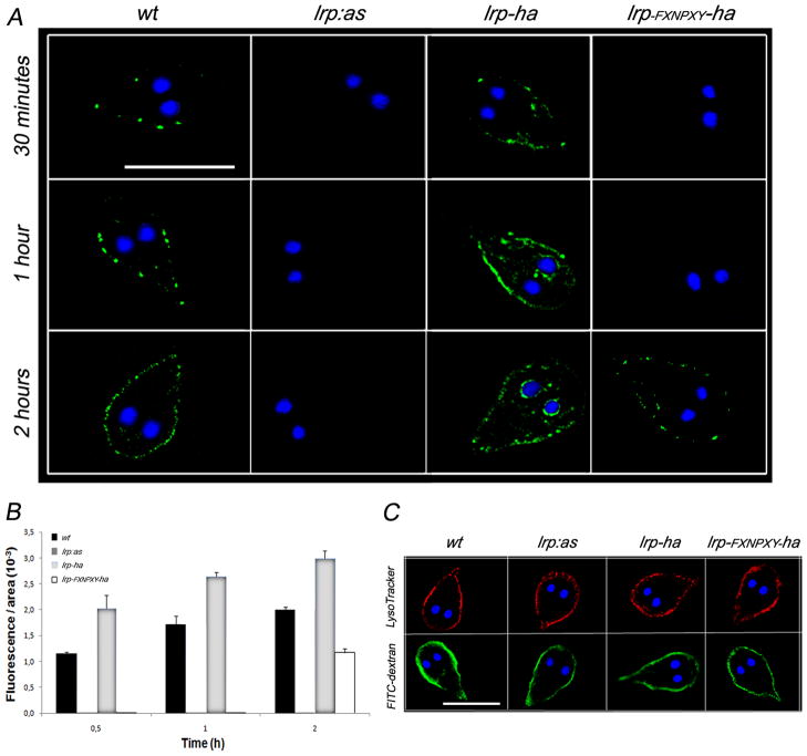
Figure 8. The endocytosis of LDL depends of the level of GlLRP expression.
(A) Immunofluoresce microscopy shows that Bodipy-LDL is endocytosed to the PVs in wild-type trophozoites (wt) but not in lrp:as trophozoites (up to 2 h). lrp-ha cells overexpressing LRP-HA show an increase of LDL internalization over time. lrp-FXNPXY-ha trophozoites show a remarkable delay in LDL endocytosis and PV delivery. The most representative effect is shown for each type of cell. Bar, 10 μm. Nuclear DNA was labeled with DAPI (blue). DAPI: 4',6-diamidino-2-phenylindole. (B) Histograms of fluorescent images (fluorescence/area × 103) show the relative amount of Bodipy-LDL internalized by parasites in control (wt) and transgenic cells (lrp-ha, lrp- FXNPXY-ha, and lrp:as). All images were equally processed; the threshold value was determined and is exclusive in each image. The results are presented here as the average (± S.D.) of ten determinations. (C) Controls adding LysoTracker Red or FITC-dextran for 30 min shows no alteration of fluid-phase endocytosis in all cell types. Bar, 10 μm.

