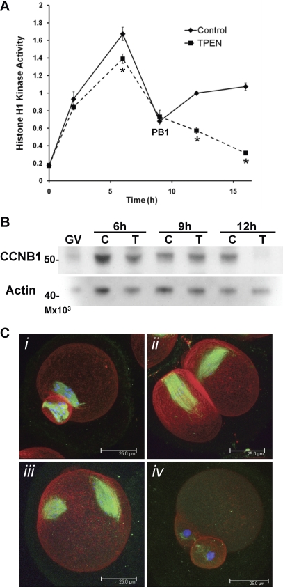
FIG. 7.
Zinc-insufficient oocytes fail to increase MPF activity following the first meiotic division. A) Kinetics of histone H1 kinase activity over the time course of IVM are shown. Values are in arbitrary units and represent densitometric analysis normalized to control oocytes matured for 12h ± SEM. Four to eleven oocytes were assayed for each data point. Asterisks indicate statistical significance according to Student t-test (P < 0.01). B) Western blot analysis for CCNB1 in oocytes matured in control (C) or TPEN (T)-containing medium for 6, 9, or 12 h. The experiment was repeated three times; a representative blot is shown. C) Three-dimensional projected confocal Z stacks of eggs stained for tubulin (green), actin (red), and DNA (blue) are shown. Following injection with CCNB1(Δ90)-EGFP cRNA during IVM in TPEN-containing medium, 26% of the injected oocytes had MII spindle-like structures (C, i and ii), while 32% did not produce polar bodies and had two spindle-like structures (C, iii), and 38% remained in MI (not shown). Uninjected oocytes matured in the presence of TPEN displayed telophase I-arrested spindles (C, iv). A total of 156 injected oocytes were analyzed. Bar = 25 μm.

