
Figure 4.
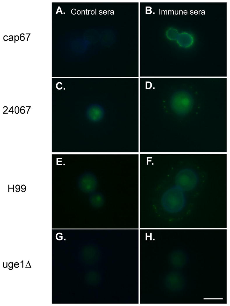
Detection of antibody responses against GalXM using indirect immunofluorescence staining. Control sera were incubated with (A) cap67, (C) 24067, (E) H99, and (G) uge1Δ. GalXM-BSA conjugate immune sera were incubated with (B) cap67, (D) 24067, (F) H99, and (H) uge1Δ. H99 is serotype A strain, and the others are serotype D strains. The blue rim around the cell body is the result of calcofluor staining. The green fluorescence in the cell body reflects autofluorescence [16]. Scale bar, 5 μm.