Fig. 2. Structure of resveratrol.
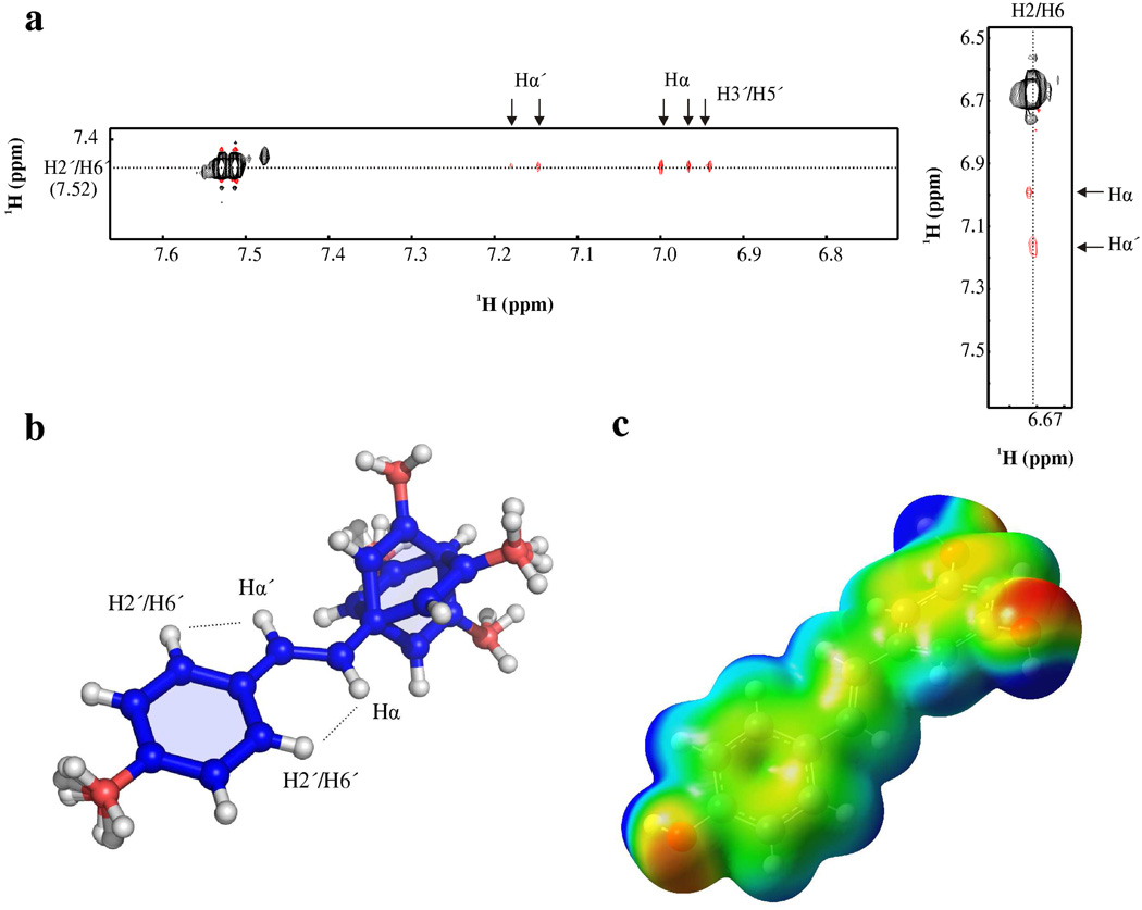
a. Two-dimensional-ROESY spectrum of resveratrol in D2O. b. Ensemble of resveratrol aligned to the olefin atoms: Hα’, Cα’, Hα, and Cα. ROEs measured between H2’/H6’ and Hα and Hα’ are drawn on the structure to illustrate that the intensity of the ROE between H2’/H6’ and Hα requires a planar orientation of phenol ring. c. Gaussian calculation from lowest energy structure of resveratrol. The electrostatic potential of resveratrol was mapped with an isovalue of 0.004 e/Å3 (−7e−2 eV, red; 7e2+ eV, blue).

