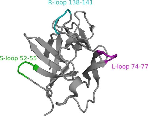
Figure 1.
NMR structure of interleukin-1β (IL1β).65 sLBTs have been incorporated into three different loops shown here in purple for the L-loop (residues 74 to 77), in green for the S-loop (residues 52 to 55) and in cyan for the R-loop (residues 138 to 141).

