Fig. 5.
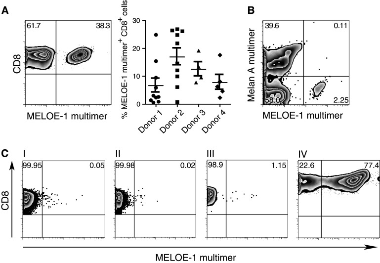
Expansion of antigen-specific T cells MELOE-136–44 after a single stimulation. a Naïve T cells from 4 different healthy HLA-A0201+ donors were stimulated with semimature DC loaded with the MELOE-1 peptide. Cultures were evaluated on day 10 using an MHC-multimer. One exemplary staining is shown (left) as well as the summary of the different parallel wells for each donor. b Expansion of T cells with different specificities in the same culture. Dendritic cells were either pulsed with the Melan-A26–35A27L peptide or the MELOE-136–44 peptide separately, washed, mixed and incubated with naïve T cells. Evaluation was performed on day 10 of the culture. Plots show live CD8+ T cells. c Enrichment of antigen-specific T cells prior to stimulation results in robust expansion similar to the kinetics seen for Melan-A-specific T cells. Naïve T cells were stained with the MELOE-1-multimer-APC (I) and subsequently coincubated with anti-APC-beads. The cells were then separated via a magnetic column into the negative fraction (II) or the positive fraction (III). The positive fraction was cultured overnight in T cell medium containing IL-7 and stimulated with MELOE-136–44-pulsed dendritic cells the next day. Expansion of the MELOE-1-specific T cell line on day 10 is shown in plot (IV)

