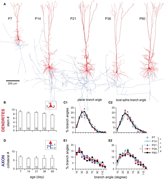
Figure A1.
The development in general structural frame of TTL5 neurons. (A) Representative 3-D model neurons reconstructed from biocytin-labeled neurons at all ages examined (the same figures as in Figure 4A; dendrites in red, axons in blue). In general, the structures of these TTL5 neurons look similar except the P7 one that was smaller and had many filopodia on its apical dendrite. Note the axon of the P60 neuron is incomplete due to faint staining. Scale bar, 200 μm. (B,C1,C2) At all ages examined, no significant changes were tested in the number of dendritic stems and the dendritic branch angles that include planer branch angle (PBA) and local spline branch angle (LSBA). (D,E1,E2) At all ages examined, no significant changes were tested in the number of collateral stems (emerged from main axon) and in the axonal PBA and LSBA.

