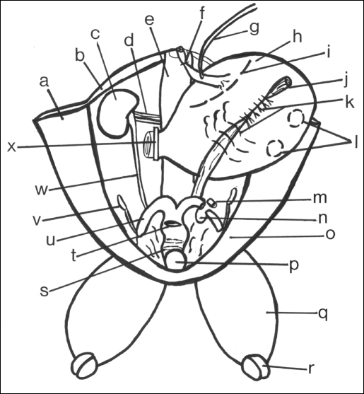
Figure 1.
Drawing of a rabbit carcass model.
- (a) Inner right abdominal wall flap
- (b) Posterior abdominal muscles
- (c) Right kidney
- (d) Right renal vessels
- (e) Spine and retroperitoneal structures
- (f) Tie anchoring esophagus to spine
- (g) Catheter through esophagus into stomach
- (h) Stomach distended with saline
- (i) Site for an electrocautery incision
- (j) Sigmoid remnant embedded in stomach
- (k) Interrupted sutures
- (l) Site for purse-string sutures
- (m) Segment of left uterine horn removed prior to anastomosis
- (n) Opening in broad ligament
- (o) Inner left abdominal wall flap
- (p) Urinary bladder
- (q) Left thigh with intact femoral vessels
- (r) Site of disarticulation of leg
- (s) Circumferential incision in body of uterus for end-to-end anastomosis
- (t) Incision site in body of uterus for running anastomosis
- (u) Right horn of uterus
- (v) Right ovary
- (w) Right ureter
- (x) Clamp across pylorus

