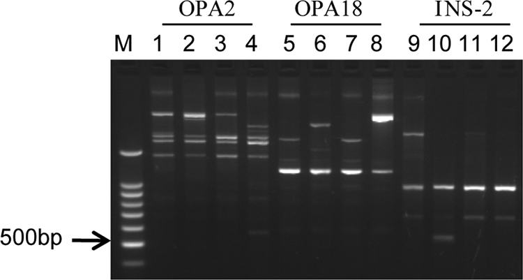
Fig. 2.

Comparison of the RAPD-PCR patterns of the clinical isolates (isolates 1 and 2), the environmental isolate, and a reference strain (M. massiliense JCM 15300T) with three different primers. Lanes 1, 5, and 9, DNA from isolate 1; lanes 2, 6, and 10, DNA from isolate 2; lanes 3, 7, and 11, DNA from the environmental isolate; lanes 4, 8, and 12, DNA from the M. massiliense reference strain; lane M, DNA size marker (100-bp ladder). RAPD-PCR patterns produced with primers OPA2 (lanes 1 to 4), OPA18 (lanes 5 to 8), and INS-2 (lanes 9 to 12) are shown.
