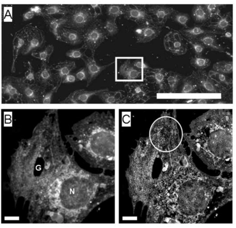
Fig. 1.

(A) Conventional deconvolution microscopy of LSECs stained with CellMask Orange, used to stain the cell membrane, showed that LSECs were intact and growing in a near- confluent monolayer. (Scale bar 20 μm). A selected cell of interest is examined at higher magnification below.
(B) The selected cells from the image above were visualized using fluorescence deconvolution.
(C) SIM image of the same cells showing improved resolution including fenestrations (circled). (Scale bar 2 μm, N nucleus, G gap).
