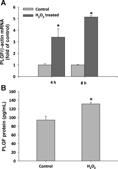
Fig. 7.
PLGF gene and protein expression is increased in response to H2O2 in primary human CASMC. A: serum-starved human coronary artery SMC (CASMC) were treated with 50 μM H2O2 for 4 and 8 h. PLGF gene expression increased to 3.4 ± 0.7-fold of control (4 h) and 5.1 ± 0.1-fold of control (8 h) (n = 3). *P < 0.05 vs. time-matched control (t-test). B: human CASMC were treated with 50 μM exogenous H2O2 for 8 h. PLGF protein levels in media increased from 91 to 131 pg/ml following H2O2 treatment (n = 3). *P < 0.05 (t-test).

