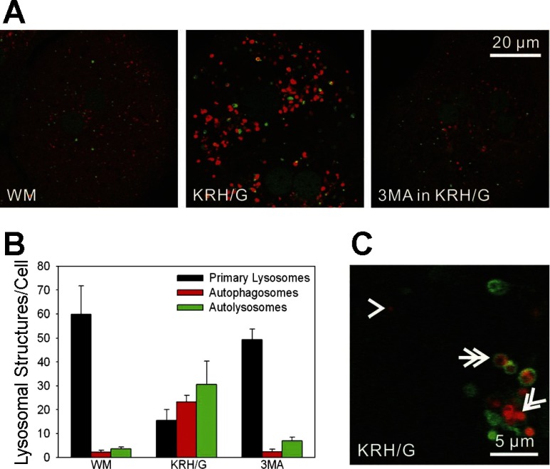
Fig. 3.
Acidification of autophagosomes during nutrient deprivation plus glucagon in GFP-LC3 hepatocytes. GFP-LC3 hepatocytes were loaded with LysoTracker Red (LTR) to label acidified vesicles, as described in materials and methods. A: GFP-LC3 hepatocytes were incubated in WM (left), KRH/G (middle), or KRH/G plus 10 mM 3MA (right) for 90 min. After incubation in WM, individual PAS were distributed throughout the cytosol, and LTR staining was mostly confined to small primary lysosomes/late endosomes. In KRH/G, LTR-labeled vesicles proliferated, often in association with GFP-LC3 fluorescence. Cells incubated in KRH/G with 3MA were indistinguishable from cells in WM. B: the numbers of primary lysosomes/acidified late endosomes, acidified autophagosomes, and autolysosomes were quantified from confocal images of GFP-LC3 hepatocytes incubated as described in A from three different hepatocyte isolations per treatment group. Differences in numbers of primary lysosomes, autophagosomes, and autolysosomes in KRH/G compared with WM and 3MA were statistically significant (P < 0.05, n = 5 cells/group). C: representative structures of a primary lysosome (arrowhead), autophagosome (double arrow), and autolysosome (double arrowhead).

