Fig. 2.
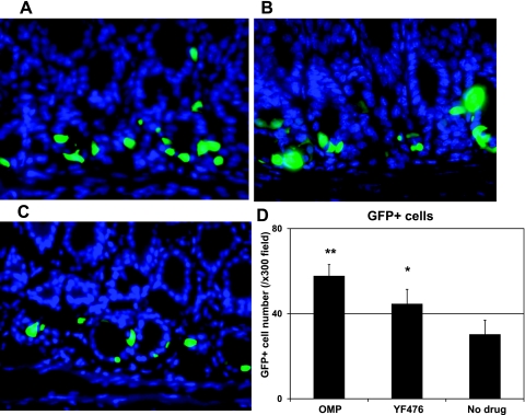
Increased intensity of GFP signals and GFP-positive cell numbers in the gastric antrum of mGAS-EGFP mice after administration of acid-suppressive reagents. A–C: merged images of GFP expression with DAPI staining in the antrum of mGAS-EGFP mice (magnification: ×600). A: mice were injected intraperitoneally with omeprazole (OMP; 5 days per week) for 2 wk. B: mice were injected intraperitoneally with gastrin/CCK2 receptor antagonist YF476 (twice per week) for 2 wk. C: mice were injected intraperitoneally with vehicle only for 2 wk. D: total numbers of GFP-positive cells in the antrum of mGAS-EGFP mice by administration with OMP or YF476 or vehicle only. The number of GFP-positive cells was expressed as the average number of positive cells counted per ×300 magnification high-power field. From each slide, cells were counted from at least 5 fields, and 3 slides (=3 mice) from each group were analyzed. The administration of either OMP or YF476 for 2 wk increased the intensity of GFP signals as well as the number of GFP(+) cells in the gastric antrum of the mGAS-EGFP mice. *P < 0.05, **P < 0.01; n = 3 for each group.

