Fig. 4.
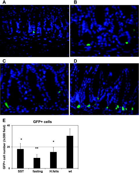
Decreased GFP signal intensity and GFP-positive cell numbers in the gastric antrum of mGAS-EGFP mice following administration of somatostatin, overnight fasting, and Helicobacter felis infection. A–D: merged image of GFP expression with DAPI staining in the antrum of mGAS-EGFP mice (magnification: ×600). A: mice were systemically infused with octreotide (long-lasting somatostatin ortholog) for 2 wk by use of osmotic minipumps. B: mice were fasted overnight. C: mice were infected with H. felis for 6 wk. D: mice without treatment (wt) or infection. E: total numbers of GFP-positive cells in the antrum of mGAS-EGFP mice by systemic infusion with the somatostatin ortholog octreotide (SST) for 2 wk, overnight fasting, and H. felis infection for 6 wk. The number of GFP-positive cells were expressed as the average number of positive cells counted per ×300 magnification high-power field. From each slide, cells were counted from at least 5 fields, and 3 slides (=3 mice) from each group were analyzed. The systemic infusion with somatostatin for 2 wk or overnight fasting or H. felis infection for 6 wk decreased the intensity of GFP signals as well as the number of GFP(+) cells in the gastric antrum of the mGAS-EGFP mice. *P < 0.05, **P < 0.01; n = 3 for each group.

