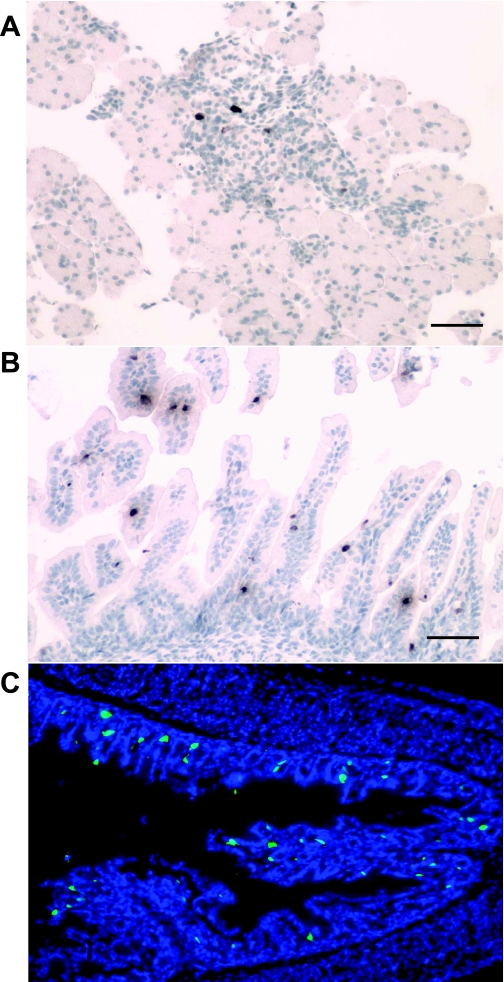
Fig. 7.
GFP expression in the fetal and neonatal organs of mGAS-EGFP mice. A and B: immunohistochemical staining with anti-GFP antibody in the fetal pancreas (A) and small intestinal villi (B) of mGAS-EGFP mice at 18.5 days postconception (dpc). C: merged image of GFP expression with DAPI staining in the gastric antrum of neonatal mGAS-EGFP mice at 2 days after birth (magnification for A–C: ×200; scale bar for A and B: 50 μm). GFP(+) cells were detected in the pancreatic islets and the small intestinal villi of the fetus of mGAS-EGFP mice at 18.5 dpc, whereas GFP(+) cells appeared in the gastric antrum of neonatal mGAS-EGFP mice at 2 days after birth. Of note, GFP(+) cells in the neonatal antrum were located at not only the base of the antral glands but also higher up in the middle to the top third of the glands.

