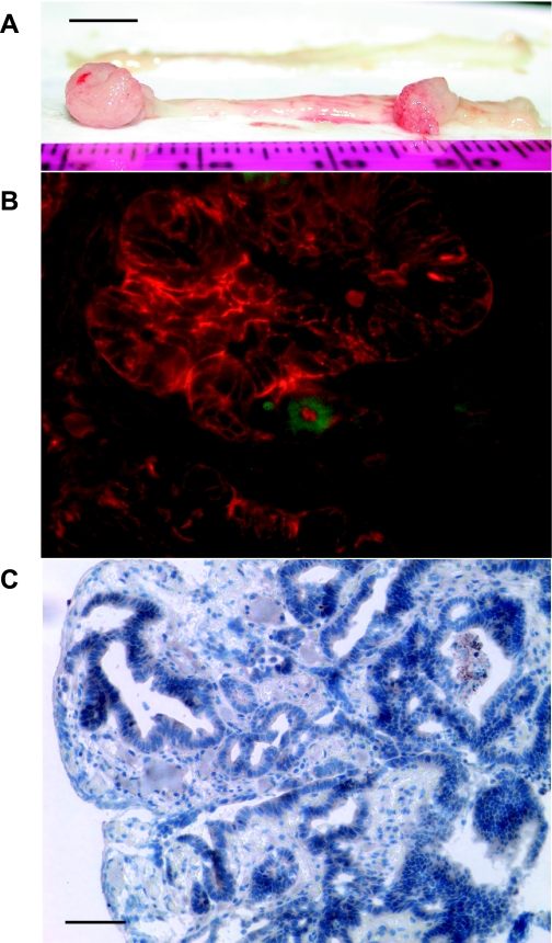
Fig. 9.
GFP expression in the cancer cells of the colon tumors induced by administration with azoxymethane (AOM) and dextran sodium sulfate (DSS) in hyperprogastrinemic hGAS/mGAS-EGFP double-transgenic mice. A: macroimage of the colorectal tumors of hGAS/mGAS-EGFP double-transgenic mice after 4 mo of administration with AOM and DSS (scale bar: 5 mm). B: merged image of GFP expression with fluorescence immunostaining with anti-EpCAM antibody followed by Texas red-conjugated anti-rat secondary antibody in the colorectal tumors of hGAS/mGAS-EGFP mice after 4 mo of administration with AOM and DSS (magnification: ×600). C: immunohistochemistry with anti-GFP antibody in the colon tumors of hGAS/mGAS-EGFP mice after 4 mo of administration with AOM and DSS (scale bar: 50 μm, magnification: ×200). Most of the GFP(+) cells were detected in the neoplastic colonic glands, and they were EpCAM positive, which indicated they were epithelial cancer cells.

