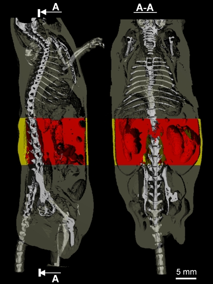
Fig 1.
A sagittal (left panel) and coronal (right panel) view of a mouse body scanned by in vivo microcomputed tomography. The skeleton was superimposed upon the adipose tissue (gray) to allow for greater spatial clarity. The abdominal VOI, in which the separation between subcutaneous (yellow) and visceral (red) fat was performed, was defined by precise skeletal landmarks.

