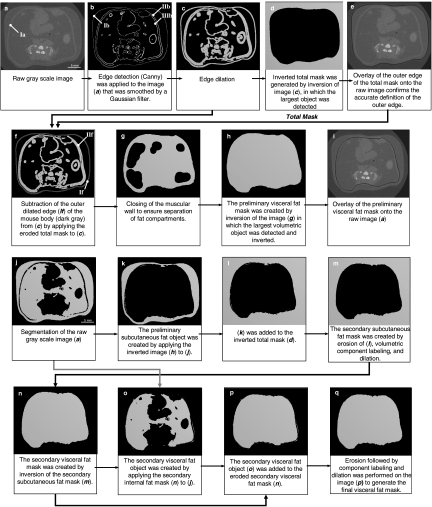
Fig 2.
Identification of the outer edge (a–e) and separation of visceral and subcutaneous fat (f–i). The contours Ia and IIf represent the abdominal muscular wall. Ib is the outer edge of the mouse body. IIb defines the interface of the muscular wall and VAT. IIIb defines the interface of the muscular wall and SAT. In images j–m and n–q, the subcutaneous and visceral fat mask were generated, respectively. Objects in black represent background.

