Fig. 6.
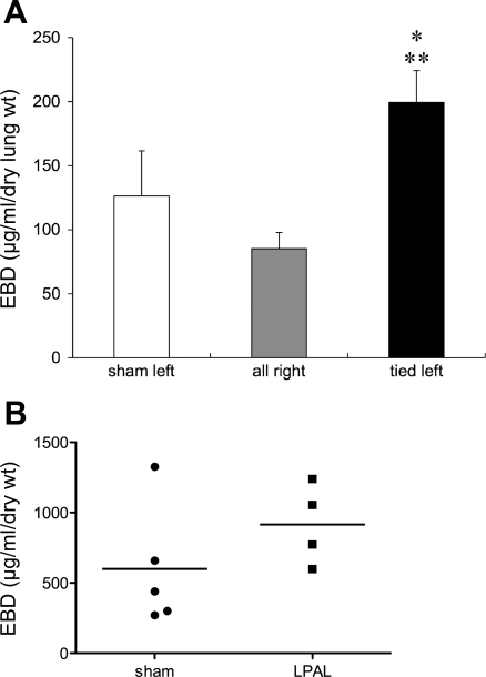
A: lung vascular permeability measured as Evans blue dye (EBD; μg·ml−1·g dry lung wt−1) concentration in left lungs from sham-operated rats, LPAL-tied left lungs, and right lungs of sham-operated and LPAL rats (n = 5 rats/group). Sham left lungs did not differ from right lungs, yet LPAL-tied left lungs showed significantly greater extravasation than sham left lungs (*P < 0.05) and right lungs (**P < 0.01). B: Evans blue dye extravasation in the trachea of sham-operated and LPAL rats. Each point represents an individual rat. Differences between sham-operated and LPAL rats did not reach statistical significance (P = 0.079).

