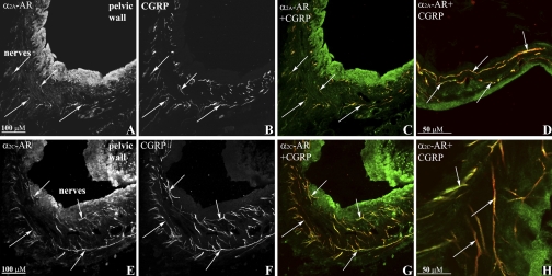
Fig. 6.
Immunofluorescence double-labeling of renal tissue with antibodies against α2A-adrenoceptors (AR), α2C-AR (green) and calcitonin gene-related peptide (CGRP; red) shows α2A-AR-immunoreactive (ir) fibers (A) and α2C-AR-ir fibers (E) close or on CGRP-ir sensory nerve fibers (B, C, and F, G, respectively) (colocalization yellow, arrows) in the renal pelvic wall. Higher magnification showed α2A-AR-ir (green, D) and α2C-AR-ir (green, H) on CGRP-ir fibers (red), colocalization yellow (arrows).

