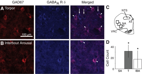
Fig. 5.
GAD67 and GABAAR δ-immunoreactivity in the VRC during hibernation. A and B: colocalization of GAD67 and GABAAR δ-immunoreactivity in neurons dorsolateral to the VRC in T and IBA. Some, but not all, GABAAR δ-immunoreactive neurons were immunopositive for GAD67. Double-labeled neurons are indicated by arrowheads in the merged image. Arrows indicate cells positive for GABAAR δ-subunit but not GAD67. C: 3 regions dorsolateral to the VRC were analyzed in each of 8 coronal sections through the VRC (P1, P2, and P3, see Supplemental Fig. S2). D: no double-labeled neurons were found in SA animals. Number of double-labeled neurons in T and IBA animals was significantly greater than SA. *P < 0.05, different than SA.

