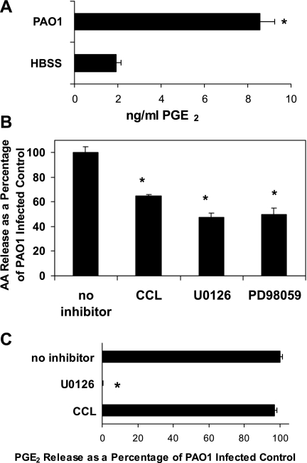
Fig. 1.
Effect of signaling kinase pathway inhibitors on PAO1-induced release of arachidonic acid (AA) and PGE2 from lung epithelial cells. A: amount of PGE2 (ng/ml) secreted from A549 cells measured by enzyme immunoassay in the presence or absence of PAO1 infection. B: relative amount of AA released by PAO1-infected A549 cells following a 1-h pretreatment with 5 μM Chelerythrine chloride (CCL), 40 μM U0126, or 40 μM PD98059 compared with vehicle control. Vehicle control (0.1% DMSO) represents the amount of AA released upon infection with PAO1 in the absence of inhibitors and is set at 100%. C: relative amount of PGE2 secreted by PAO1-infected A549 cells preincubated for 1 h before infection with either 5 μM CCL or 40 μM U0126 compared with vehicle control. Vehicle control (0.1% DMSO) represents the amount of PGE2 secreted upon infection with PAO1 in the absence of inhibitors and is set at 100%. *P < 0.05, statistically significant difference compared with control using the Student's t-test. Each data point represents an average of at least 3 separate wells. Evaluation of each inhibitor was performed in at least 3 internally controlled experiments yielding similar results.

