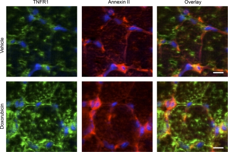
Fig. 5.
TNFR1 localization after doxorubicin exposure. Panels show representative confocal images of transverse sections from diaphragms of mice treated with vehicle (top) or doxorubicin (bottom); muscle was collected and processed 72 h postinjection. Sections were stained for anti-TNFR1 (green) and counterstained with anti-annexin II (red) and DAPI (blue), a nuclear stain. Scale bar = 10 μm.

