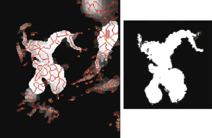
Fig 3.
Results of the watershed segmentation (left), thresholded lightly to remove some soft tissue and background segments. The lines indicate the segment boundaries. The segment-grown image (right) shows the detected biliary structure. Some noise, artifacts, and other organs were removed through the structure detection stage.

