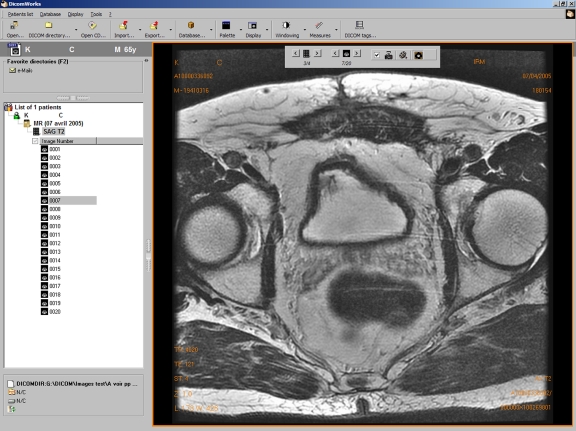
Fig 2.
Screenshot of the main screen of version 1.3.5 with a study opened (axial T2 weighted sequence of a prostate MRI examination). On the left part of the screen, a direct access to favorite directories of DICOM images is possible (blue folder), whereas the current directory is sorted in patients, studies, series, and images. All levels are expandable (such as here at the image level). On the right part of the screen, a maximum of the screen surface is dedicated to the image. Patient and image data are overlaid. A simple toolbar allows easy navigation through the series and images. Window leveling, pan, and zoom are performed using the mouse, and the mouse wheel allows scrolling in the series. Important functions (measures, screen splitting, etc...) are accessible from the toolbar.

