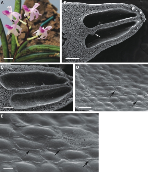
Fig. 13.
Inflorescence and spur of Stereochilus dalatensis; (B–E) scanning electron micrographs. (A) Zygomorphic, pink and rose-coloured flowers with saccate spurs (arrows). Scale bar = 5 mm. (B) Bisected spur showing two loculi (lumina) separated by a median, longitudinal septum with proximal protuberance (arrow). Scale bar = 0·5 mm. (C) Detail of the two spur lumina lined with glabrous epidermis. Scale bar = 200 µm. (D) Epidermal cells showing distension of cuticle at cell junctions (arrows). Scale bar = 50 µm. (E) Secretory epidermis showing distension of cuticle (arrows) and nectar residues. Scale bar = 10 µm. Abbreviations: see Fig. 1.

