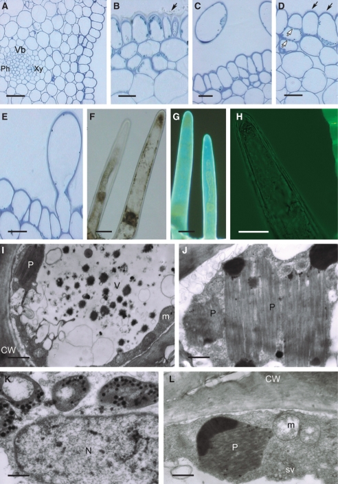
Fig. 8.
Histology and ultrastructure of spur of Papilionanthe vandarum: (A–H) light micrographs; (I–L) transmission electron micrographs. (A) Section of spur wall showing epidermis, underlying tissues and vascular bundle. Scale bar = 50 µm. (B) Glabrous epidermis cells with nectar residues upon its surface (arrow). (C) Transverse section through epidermis and secretory hairs showing parietal cytoplasm with plastids. (D) Epidermal cells with striated cuticle (solid arrows). Pit-fields are present in epidermal and subepidermal cells (open arrows). (E) Epidermis with secretory, unicellular hair cut longitudinally and showing smooth cuticle. (B–E) Scale bars = 20 µm. (F) Unicellular, secretory hairs with large nuclei and numerous plastids in parietal cytoplasm. (G) Hairs showing autofluorescence and that plastids lack chlorophyll. (F, G) Scale bars = 25 µm. (H) Secretory hair following treatment with auramine O, yet showing no fluorescence. Scale bar = 25 µm. (I) Transverse section of hair showing cytoplasm with mitochondria and plastids. Vacuole contains vesicles and dark precipitates. Scale bar = 4 µm. (J) Plastid with numerous, dense lamellae and osmiophilic regions. Scale bar =1·5 µm. (K) Detail of cytoplasm with nucleus and chromoplasts. Scale bar = 1 µm. (L) Parietal cytoplasm with plastid, mitochondria and secretory vesicles. Scale bar = 1·5 µm. Abbreviations: see Fig. 1.

