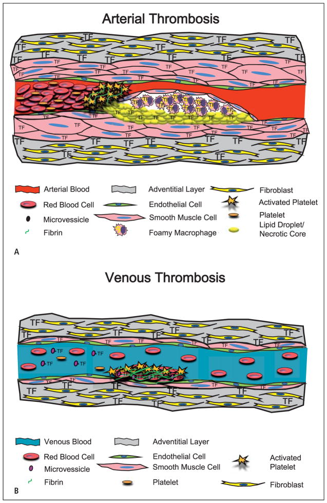
Figure 1. Models showing the cellular sources of TF that contributes to arterial and venous thrombosis.
A) Arterial thrombosis. The primary trigger of arterial thrombosis in a diseased vessel is the rupture of an atherosclerotic plaque. This involves disruption of the fibrous SMC cap exposing blood to TF in the plaque. The resultant clot is mainly composed of platelets with lower levels of cross-linking fibrin, and is referred to as a “white clot”. B) Venous thrombosis. Venous thrombosis occurs on a relatively undisturbed endothelial layer. Venous thrombosis may be triggered by circulating TF associated with MVs. The clot is mainly composed of fibrin with platelets and trapped red blood cells and is referred to as a “red clot”.

