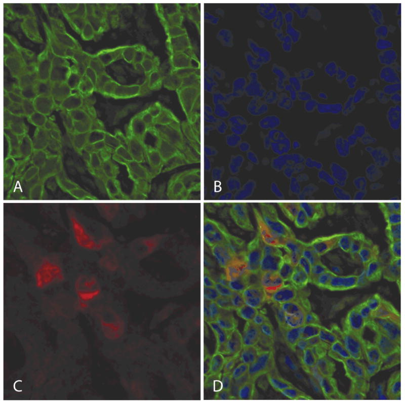
Figure 1.

Protein expression of ribonucleotide reductase M2 (RRM2) was determined using an automated in situ quantitative measurement of protein analysis, automated quantitative immunohistochemistry on the basis of immunofluorescence.
a. Target compartments were localized using a fluorescently tagged Alexa Fluor 555, rabbit anti-cytokeratin antibody (green). b. 4,6-Diamidino-2-phenylindole was added to visualize nuclei (blue). c. RRM2 was visualized with Alexa Fluor 488-tyramide, human anti-RRM2 (red).
d. A three color overlay shows localization of RRM2 to the cytoplasm.
