Fig. 2.
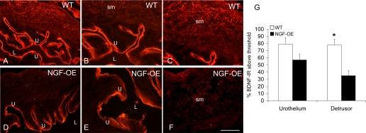
A–F: BDNF immunoreactivity (IR) in urothelium (U; A, B, D, E) and detrusor smooth muscle (sm) (A, C, D, F) of littermate WT (A–C) and NGF-OE (D–F) mice. Comparable BDNF IR was present in urothelial cells of WT and NGF-OE urothelium (A, B, D, E). BDNF-IR expression was reduced in detrusor sm of NGF-OE mice (D, F) compared with detrusor sm of WT mice (A, C). L, lumen. Calibration bar: 50 μm (B, C, E, F), 125 μm (A, D). G: summary histogram of BDNF expression in U and detrusor sm in WT and NGF-OE mice. Values are means ± SE (n = 5–7). *P ≤ 0.01.

