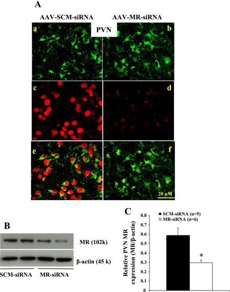
Fig. 4.
Immunohistochemical and Western blot analyses of MR protein expression in the PVN from rats receiving icv injections of SCM-siRNA or MR-siRNA. A: adenovirus-mediated delivery of RNA interference effectively silences MR (red in the nucleus) expression in the PVN. Green in the cytoplasm represents GFP, a marker for virus transfection. Images in a–d are merged in e and f. B: representative Western blots of MR and β-actin. C: the results of Western blot analysis represent the change in MR protein expression, which was normalized with β-actin in the PVN. *P < 0.05 compared with SCM-siRNA treatment.

