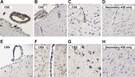
Fig. 5.
Immunohistochemistry of CSE (A–D) and CBS (E–H) in newborn pig cerebral cortex detected by the avidin-biotin-peroxidase complex technique. A and B: CSE-positive (brown) pial arterioles on the cerebral surface. B and C: CSE-positive (brown) penetrating cerebral vessels. E: CBS-negative pial arteriole on the cerebral cortical surface and an adjacent CBS-negative venule. F: CBS-negative parenchymal vessel. F and G: abundant CBS-positive (brown) neurons and astrocytes, including pyramidal cells (G). D and H: control slices treated with secondary antibody only. Counterstaining: hematoxylin. A/B, antibody.

