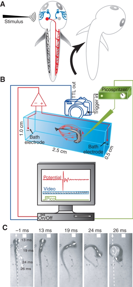
Fig. 1.
Zebrafish escape circuit and experimental setup. (A) A simplified diagram of the Mauthner-mediated escape circuit. Mechanosensory stimuli activate trigeminal sensory neurons (blue) that synapse onto the ipsilateral Mauthner neuron (red). The Mauthner cell axon crosses the midline and activates the axial motor neurons and respective muscles on the contralateral side. (B) The setup for recording electric field potentials generated during escape behavior. Behavior was evoked by applying a jet of water to the head and was recorded by high-speed videography (1000 frames s–1). Simultaneously, electric field potentials (red ovals surrounding behaving animal in chamber) were measured using a differential amplifier. Electric field recordings and videos of behavior (red and blue components on computer display) were digitally synchronized with the onset of the Picospritzer water jet (green component on display). This ensured time-locking of the field potential, escape behavior and water jet pulse. (C) Changes in head trajectory during escape behavior were measured as the angle between the original body axis (–1 ms, just prior to the first detectable movement) and lines drawn through the head at subsequent time points (13, 19, 24 and 26 ms later). In this example, the maximal change in head trajectory occurred 26 ms after the first detectable movement. Angular velocity was calculated by measuring the change in head trajectory as a function of frame number, with each frame corresponding to 1 ms.

